Echocardiograms for Pets
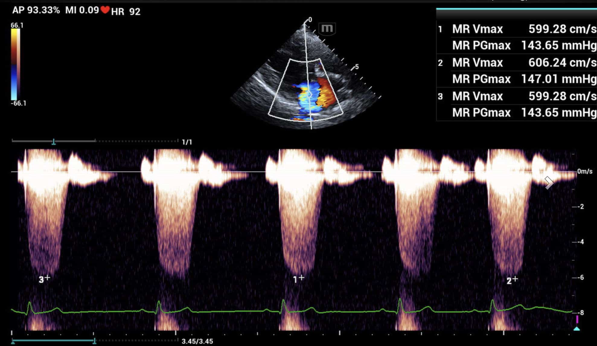
By adding mobile echocardiograms in your clinic, you offer pet parents a more convenient and accessible diagnostic option. Keeping patients in your clinic, reduces the need to travel long distances to specialty care facilities, while reducing wait times and stress for both the pet and pet parent.
Clinical Reasons to Prioritize Echocardiography
Diagnose Cardiac Conditions Early
Detect structural abnormalities like valvular disease, cardiomyopathy, pericardial effusion, or congenital defects
Differentiate between cardiac and non-cardiac causes of symptoms (e.g., cough, collapse, exercise intolerance)
Assess severity and progression before outward signs become obvious
Guide Treatment Plans
Tailor medication protocols based on specific chamber enlargement, contractility, and pressure estimates
Avoid empiric or potentially harmful cardiac medications without a confirmed diagnosis
Monitor response to therapy over time
Support Pre-Anesthetic Screening
Identify occult heart disease in senior patients
or at-risk breeds before anesthesia or surgery
Reduce anesthesia-related complications by
Understanding cardiovascular risk
Breed-Specific Screening
Cavalier King Charles Spaniels (mitral valve disease)
Golden Retrievers (Diet-related DCM)
Dobermans (DCM)
Boxers (arrhythmogenic right ventricular cardiomyopathy)
Since some pets show no clear symptoms until cardiac disease is advanced, early imaging can make a significant difference in treatment and long-term health. Echocardiography provides both pet owners and veterinarians with the important insight needed to diagnose, monitor, and manage heart conditions effectively.
Understanding Echocardiograms: What They Are and Why They Matter
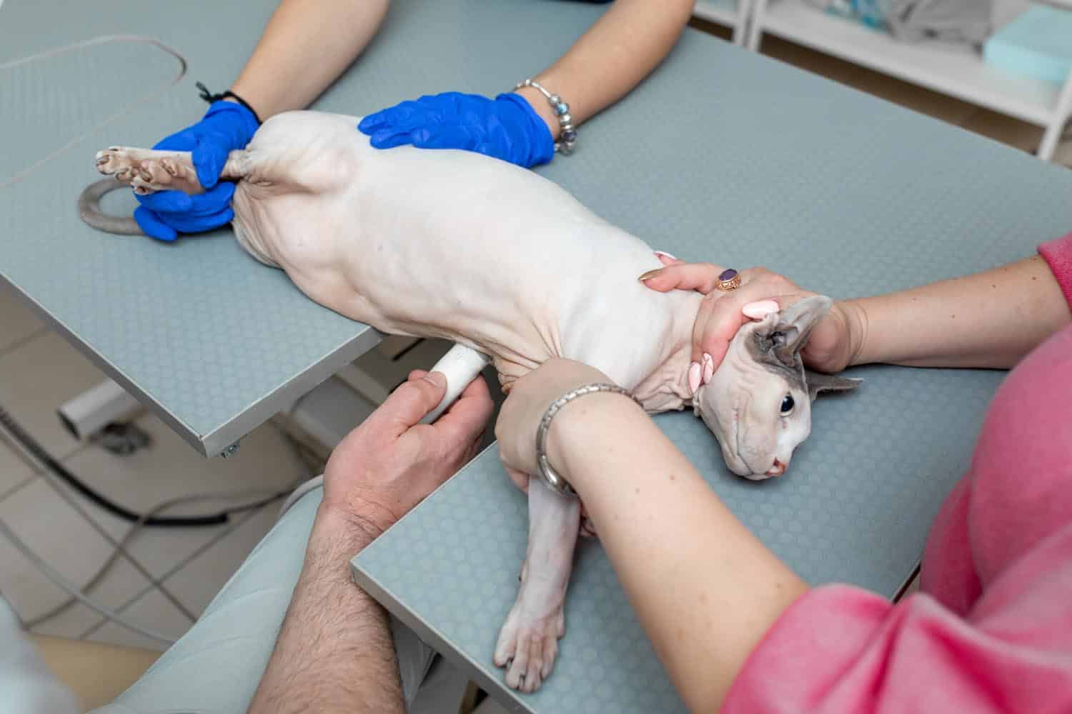
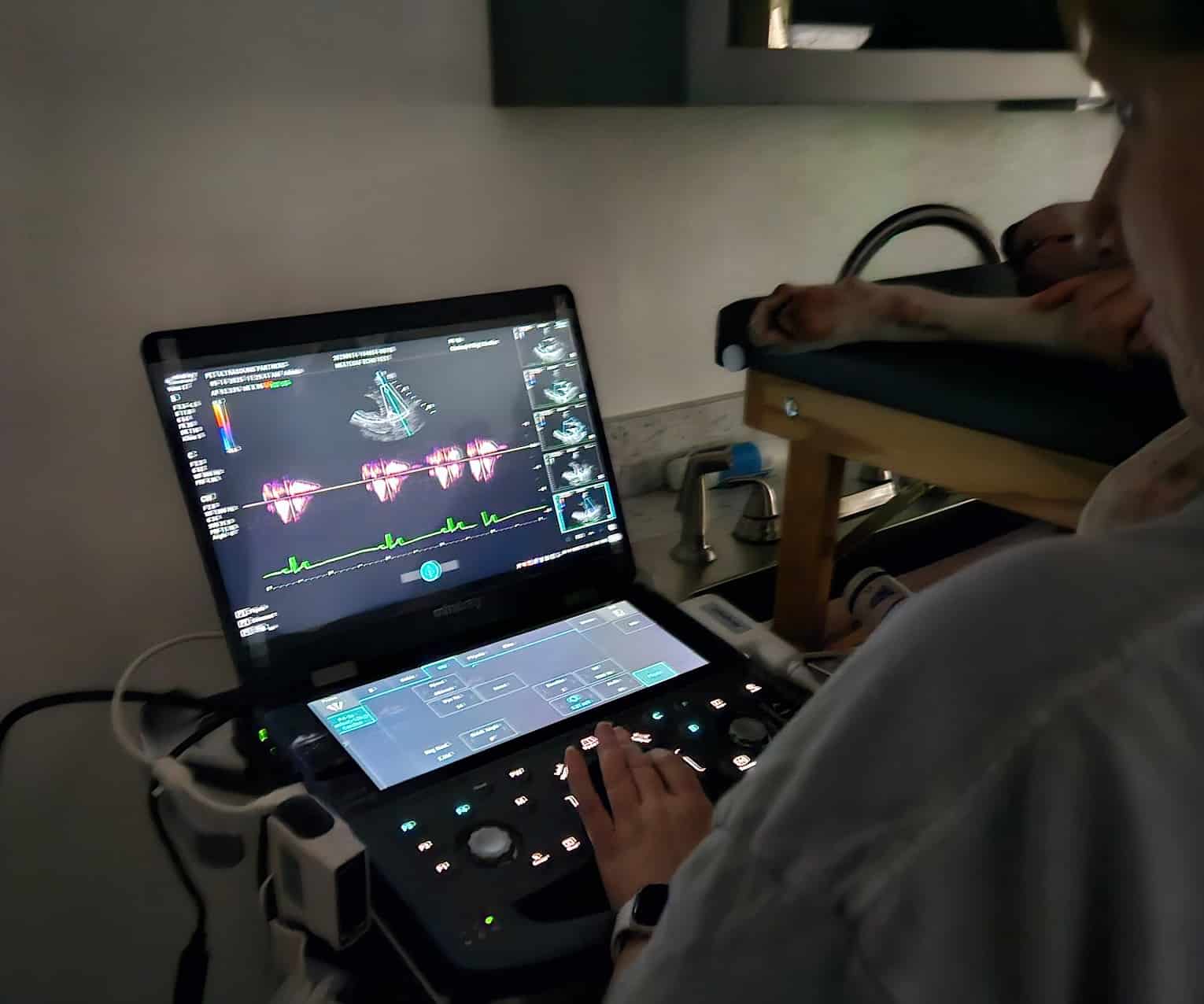
A veterinary echocardiogram is a safe, non-invasive ultrasound that provides real-time images of a pet’s heart, allowing veterinarians to assess its structure, motion, and blood flow with precision. By using a handheld transducer on a shaved area of the chest, this procedure captures dynamic visuals of the heart’s chambers and valves—offering more detailed insights than X-rays. It’s typically painless and well-tolerated by both dogs and cats.
Echocardiograms are especially valuable for diagnosing early or hidden cardiac conditions. They are commonly recommended when a veterinarian detects a murmur, suspects heart disease, or wants to evaluate breeds known to be at risk. Some of the key conditions detected include:
Dilated Cardiomyopathy (DCM)
Enlargement and weakening of the heart muscle
Congenital Defects
Structural issues present at birth, such as septal defects or PDA
Hypertrophic Cardiomyopathy (HCM)
Thickening of the heart muscle, impairing relaxation
Pericardial Effusion
Fluid buildup around the heart that may restrict its function
Valve Disorders
Leaky or stiff valves causing murmurs and disrupted blood flow
Since some pets show no clear symptoms until cardiac disease is advanced, early imaging can make a significant difference in treatment and long-term health. Echocardiography provides both pet owners and veterinarians with the important insight needed to diagnose, monitor, and manage heart conditions effectively.
How Echocardiograms Help Veterinarians and Pets
Echocardiograms provide veterinarians with detailed, real-time images of a pet’s heart, allowing for accurate diagnosis, tailored treatment, and effective monitoring. Through the mobile services, clinics gain access to this advanced cardiac imaging right at their facility—without requiring pet owners to seek out separate referrals. For pet parents, it brings clarity and comfort knowing their veterinarian has precise, visual insights to work from.
Signs That May Prompt an Echocardiogram
Veterinarians may recommend this diagnostic test if a pet shows any of the following signs:
Coughing or shortness of breath
Audible murmurs during a routine exam
Decreased stamina or fainting episodes
Swelling in the abdomen or limbs
Irregular heartbeats
Even when no obvious symptoms are present, an echocardiogram may be used for early screening, especially in high-risk patients.
Preventative and Routine Screening Uses
Echocardiograms are often recommended for:
Senior pets
as part of regular wellness checks
Pre-anesthetic evaluations
for pets with murmurs or cardiac risk factors
Breeds predisposed to heart disease
such as: Boxers, Doberman Pinschers, Cavalier King Charles Spaniels, Maine Coon cats
Monitoring known heart conditions
to track progression or adjust treatment
Diagnostic Ultrasound Delivered
Right to Your Clinic
Pet Ultrasound Partners provides a mobile imaging solution that brings echocardiograms directly to veterinary clinics in Joplin, MO and surrounding areas. This model eliminates the need for external referrals, keeping pets in the comfort of their regular clinic and allowing veterinarians to offer advanced cardiac diagnostics on-site.
Key Benefits of Mobile Services
Convenience at Your Clinic
Pet Ultrasound Partners delivers advanced cardiac imaging directly to veterinary clinics, reducing the need for outside referrals or travel.
Less Stress for Pets
Exams are done in a familiar setting, keeping pets calmer—especially helpful for seniors and anxious patients.
Imaging You Can Rely On
Experienced sonographers use high-resolution ultrasound to capture detailed cardiac images that support accurate diagnosis.
Quick, Collaborative Results for Better Workflow and Care
Ideal for patients with limited mobility, our mobile service streamlines diagnostics while keeping care in-house. Reports are shared directly with the referring veterinarian to support prompt treatment decisions and maintain continuity of care.
What to Expect During a Pet Echocardiogram
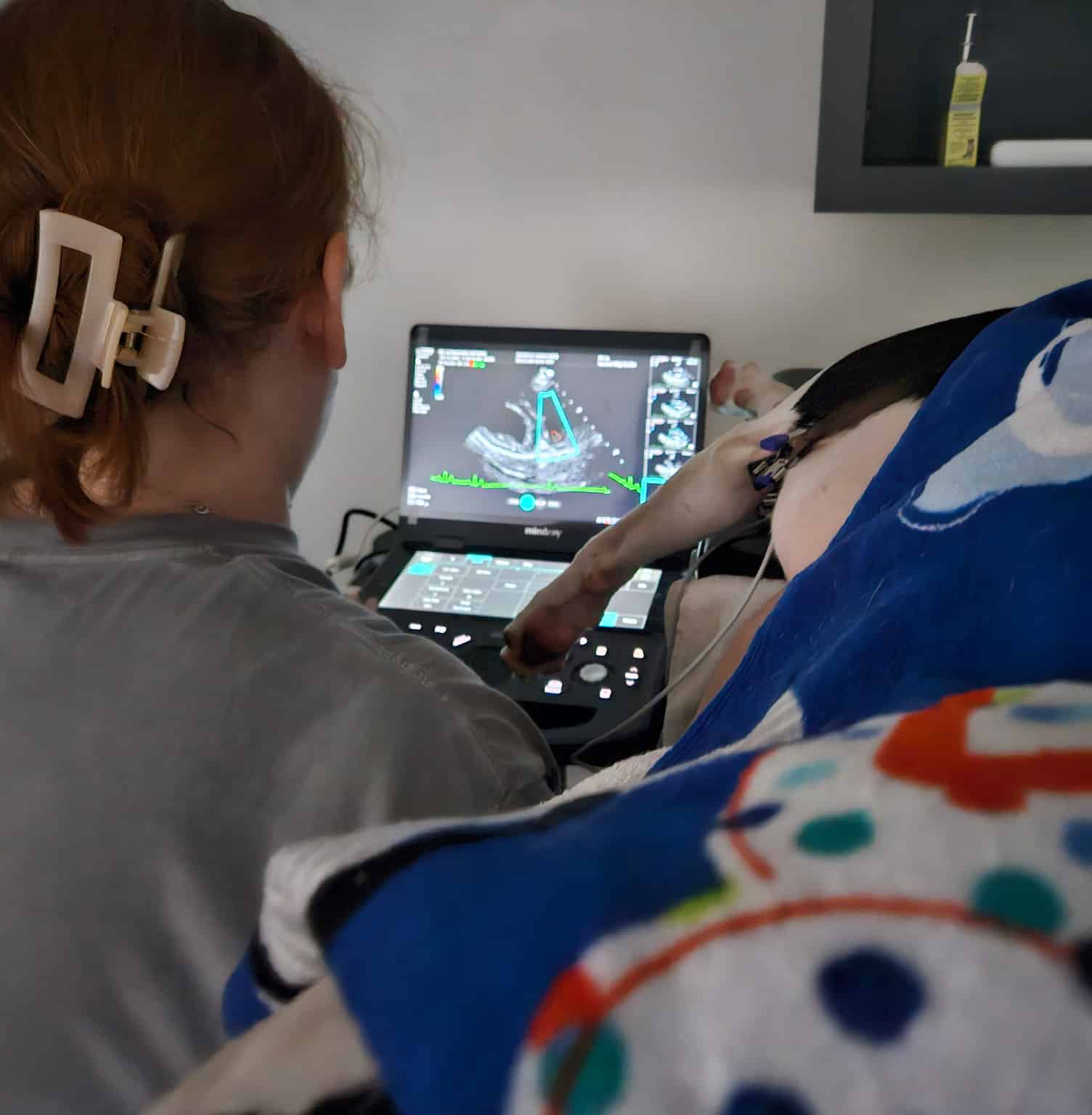
Understanding what happens during an echocardiogram can ease any concerns pet owners or clinic teams may have. The process is straightforward, non-invasive, and well-tolerated by most pets. Since the service is performed within the pet’s regular veterinary clinic, the experience is more relaxed and comfortable for both patients and their owners.
Here’s a step-by-step breakdown of what you can expect:
Light Sedation Recommended
With light sedation, most pets remain calm with gentle handling and minimal restraint. This is especially helpful for senior pets or those with anxiety, allowing for a stress-free, fast recovery.
Shaving for Ultrasound Contact
A small area of fur may need to be shaved to ensure proper contact between the ultrasound probe and the skin. This helps produce clear, high quality images.
20 to 30-Minute Procedure
The scan usually takes approximately 30 minutes. During this time, the sonographer captures real-time images of the heart from multiple angles. Pets stay awake and comfortable throughout the process.
Quick Reporting to Veterinarians
Once the echocardiogram is complete, the images are sent directly to a board-certified veterinary cardiologist for interpretation. A detailed report with treatment recommendations is then sent to the referring veterinarian. This supports timely decisions on treatment, medication, or follow-up care—keeping everything within the familiar clinic setting.
Serving Joplin and
Surrounding Missouri
Communities
Veterinary clinics across Joplin, MO trust Pet Ultrasound Partners to deliver high-quality mobile imaging that enhances their diagnostic offerings without the need for additional equipment or referrals. By bringing echocardiogram services directly into local clinics, more pets can receive timely, in-house cardiac evaluations.
Whether a practice is addressing a newly detected murmur or tracking the progress of an ongoing heart condition, Pet Ultrasound Partners provides dependable, on-site cardiac imaging that supports faster, more informed care.
Ready to Enhance Cardiac Care for Your Patients?
Mobile echocardiograms offer a convenient, low-stress solution for assessing pet’s heart health—eliminating the need for separate office visits or unfamiliar travel. Pet Ultrasound Partners collaborates directly with veterinary teams to deliver valuable imaging services on-site, complete with timely, accurate reporting that supports fast, informed care.
To schedule an echocardiogram or speak with our team, call (417) 622-1700, email us, or request an appointment online.
Frequently Asked Questions About Veterinary Echocardiograms
What is an echocardiogram and why is it recommended for pets?
An echocardiogram is a painless, non-invasive ultrasound exam that shows the heart’s internal structure and function. It helps determine the cause of heart murmurs, evaluate possible congenital heart defects and stage the of severity of valvular heart disease.
Is sedation required for an echocardiogram?
To ensure optimal image quality and reduce stress, sedation is strongly encouraged. Calm patients allow for more precise scans and shorter scanning times. We coordinate with your clinic to support sedation protocols in advance of each visit.
